-

Brother™ Printer Paper
SKU/REF 9770084
-

Seiko™ Thermal Printer Paper
SKU/REF 9770037
true


- Overview
- Products & Accessories
- EIFU & Resources
- Compatible PICC Catheters
- Integrated/Standalone
false
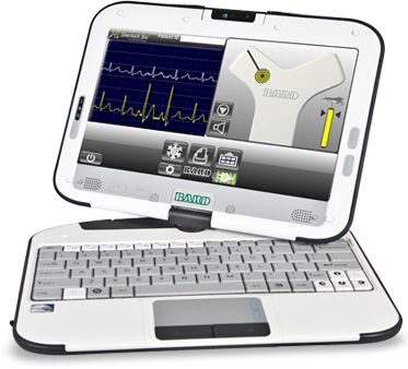
Simultaneous View of Catheter Tip Tracking and ECG
Catheter tip tracking and ECG confirmation are integrated on the same graphical display.
Static ECG baseline
Provides comparative ECG waveform for interpretation of P-wave changes and to assist in maximum P-wave identification.
Dynamic ECG waveform
Measures changes in P-wave morphology to allow the clinician to position the catheter tip in proximity to the cavoatrial junction.
Ability to maintain sterile field
Through-drape connection allows clinician to maintain maximal barrier sterile field during placement. Probe and wireless remote allow the clinician to maintain sterility while interfacing with Sherlock 3CG™ TCS.
Tip Confirmation documentation
Allows for PICC tip location to be easily documented in patient's chart or stored on the Sherlock 3CG™ TCS System.
Guideline Compliance
By helping to position the PICC tip in proximity to the cavoatrial junction, the Sherlock 3CG™ TCS System helps clinicians comply with AVA and INS guidelines for proper PICC placement.
Indicated for use as an alternative method to chest X-ray and fluoroscopy for PICC tip placement confirmation in adult patients.
Immediate confirmation of PICC tip position at the bedside and immediate release of the PICC line.
Increases placement efficiency and reduces catheter manipulations as compared to "blind" catheter placements.
Eliminates costs associated with confirmatory chest X-ray and time previously spent waiting for X-ray confirmation readings.
Eliminates confirmatory chest X-ray exposure to the patient and clinician.
Sherlock 3CG™ Tip Confirmation System (TCS) Clinical Study
Getting to the heart of the matter…
Sherlock 3CG™ Tip Confirmation System (TCS) Clinical Study
Literature
BD's collection of literature on industry and on our offerings gives you information you can use to continue striving for excellence.
Learn more
Training
BD offers training resources to help improve your clinical practices as part of our goal of advancing the world of health.
Learn more
Events
BD supports the healthcare industry with market-leading products and services that aim to improve care while lowering costs. We host and take part in events that excel in advancing the world of health™.
Learn more
Case Studies
BD promotes clinical excellence by providing various resources on best practices, clinical innovations and industry trends in healthcare.
Learn more
true
PowerPICC SOLO™ 2 Catheters with Sherlock 3CG™ TPS Stylet
| SKU/REF | Catheter Size | Lumens | Tray |
|---|---|---|---|
| 1194108D | 4 F | Single | Max Barrier |
| 1194108 | 4 F | Single | Full |
| 1295108D | 5 F | Dual | Max Barrier |
| 1295108 | 5 F | Dual | Full |
| 1295108FD | 5 F | Dual | Max Barrier (PowerPICC™ FT) |
| 1295108F | 5 F | Dual | Full (PowerPICC™ FT) |
| 1395108QD | 5 F | Triple | Max Barrier |
| 1395108Q | 5 F | Triple | Full |
| 1396108D | 6 F | Triple | Max Barrier |
| 1396108 | 6 F | Triple | Full |
PowerPICC™ Catheters with Sherlock 3CG™ TPS Stylet
| SKU/REF | Catheter Size | Lumens | Tray |
|---|---|---|---|
| 1173108D | 3 F | Single | Max Barrier (PowerPICC™ SV) |
| 1173108 | 3 F | Single | Full (PowerPICC™ SV) |
| 1174108D | 4 F | Single | Max Barrier |
| 1174108 | 4 F | Single | Full |
| 1274108D | 4 F | Dual | Max Barrier (PowerPICC™ SV) |
| 1274108 | 4 F | Dual | Full (PowerPICC™ SV) |
| 1175108D | 5 F | Single | Max Barrier |
| 1175108 | 5 F | Single | Full |
| 1275108D | 5 F | Dual | Max Barrier |
| 1275108 | 5 F | Dual | Full |
| 1275108FD | 5 F | Dual | Max Barrier (PowerPICC™ FT) |
| 1275108F | 5 F | Dual | Full (PowerPICC™ FT) |
| 1385108QD | 5 F | Triple | Max Barrier |
| 1385108Q | 5 F | Triple | Full |
| 1386108D | 6 F | Triple | Max Barrier |
| 1386108 | 6 F | Triple | Full |

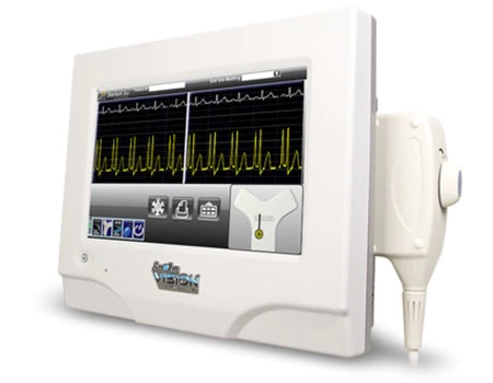
The Site-Rite Vision® Ultrasound System
Sherlock 3CG™ TCS simplifies the PICC insertion process
By combining PICC tip placement technology into select Site~Rite™ Ultrasound systems:
- Evaluate and assess patient vasculature
- Select and access the appropriate vessel
- Use Sherlock 3CG™ TCS tip tracking technology to locate and navigate the catheter tip to the SVC
- Use Sherlock 3CG™ TCS ECG technology to distinguish changes in P-wave amplitude
- Confirm catheter tip placement at the CAJ without the need for a confirmatory chest X-ray
- DICOM or printed documentation

Sherlock 3CG™ TCS is available as a separate standalone system
For customers not using the Site~Rite Vision® Ultrasound System. This device provides the same great benefits of the integrated system but provides flexibility for use across multiple imaging platforms.
false
true